


Have you noticed recently that your skin is covered in dark spots or has an uneven coloration? Then it is possible that you are suffering from hyperpigmentation, a general skin condition that results from a higher-than-normal concentration of melanin pigment in one’s skin. While hyperpigmentation can be aesthetically displeasing, many forms of hyperpigmentation are harmless – but is it possible that your form of hyperpigmentation is caused by something more sinister? We want to dedicate today’s post to differentiating some of the most common forms of hyperpigmentation, and what you can do to fade these dark spots.
Melasma
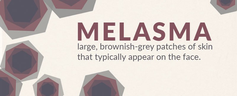
Melasma is a condition characterized by grayish-brown patches of skin that appear on sun-exposed parts of the body (face, neck, arms, etc.). Women with darker complexions and who are of child-bearing age most commonly suffer from melasma, particularly women who are of Asian, Latino, or Caribbean descent. However, it is possible for people of all genders and ethnicities to suffer from melasma. Despite its prevalence amongst Asian and Hispanic populations, most individuals will not be diagnosed with melasma until after they reach puberty. Melasma is often divided into three subcategories: malar (on the cheek), centrofacial (the nose, cheeks, and the forehead), and mandibular (along the jawbone). It is believed that melasma can be induced by factors including pregnancy, hormone therapy and use of oral contraceptives, use of certain cosmetics, genetic and racial makeup, and sun exposure. There is no known cure for melasma, though sufferers are usually treated by eliminating as many potential aggravating factors as possible. Additionally, those with melasma will likely employ lightening agents like azelaic acid and hydroquinone, in addition to a powerful broad-spectrum sunscreen. Many individuals typically see a fading in their hyperpigmentation when they avoid sun exposure. For more severe cases, chemical peels, laser therapy, and dermabrasion may be employed (Indian Journal of Dermatology, Indian Journal of Dermatology).
Melanoma
Among the most dangerous forms of hyperpigmentation (and the most dangerous form of skin cancer) is melanoma. Melanoma occurs when skin cells suffer from unrepaired DNA damage, which in turn causes genetic mutations that cause skin cells to multiply rapidly and create malignant cancerous tumors. The reason for melanoma’s dark pigmentation is partially due to tumors originating in basal layer melanocytes (pigment-producing cells), although certain melanomous moles may deviate from their typical black or brown hue and be pink, purple, red, blue, white, or flesh-colored. Intense UV exposure (typically followed by sunburn) is a major factor in the pathogenesis of melanoma, a factor which is further aggravated in individuals who have a genetic predisposition for this cancer. Having many moles or several large moles, having a family history of unusual moles or melanoma, having fair skin, and having a weak immune system have all been linked with a higher probability of developing melanoma, among other factors. As long as melanoma is caught fairly early, there is a good chance that one can be cured of the condition (almost a 100% chance). Nevertheless, melanoma is the most lethal form of skin cancer and affects 135,000 people in the United States per year (Skin Cancer Foundation, American Academy of Dermatology, National Cancer Institute).
The Skin Cancer Foundation recommends following the ABCDE’s of melanoma (and visiting your dermatologist!) to determine if your hyperpigmentation is benign or potentially melanomous:
- A for Asymmetry – Benign moles are usually symmetrical, whereas cancerous moles typically are asymmetrical.
- B is for Border – Benign moles have even, smooth borders, whereas melanomous moles have jagged, uneven borders.
- C is for Color – Benign moles tend to have a uniform color, whereas malignant moles are multi-colored.
- D is for Diameter – Benign moles are usually less than 6 mm in diameter, whereas malignant moles are 6 mm or greater in diameter. Even if your mole is less than 6 mm, you should have it looked at by a dermatologist, as many early-stage melanomous moles may be smaller than 6 mm.
- E is for Evolving – Benign moles tend to stay the same shape, size, and color as you age, whereas malignant moles often change their appearance over time.
Post-inflammatory Hyperpigmentation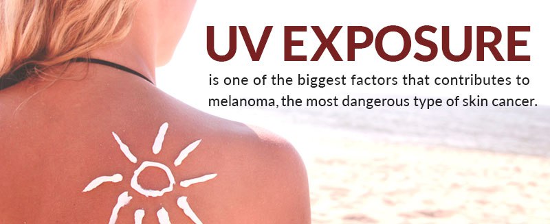
Post-inflammatory hyperpigmentation (PIH) is among the most common forms of hyperpigmentation, especially for those with darker complexions. PIH typically results after the skin undergoes some sort of inflammation or cutaneous trauma, such as a burn, cut, acne breakout, etc. In response, the body either overproduces or poorly disperses melanin pigments throughout your skin. Certain inflammatory mediators (like prostanoids and cytokines) and reactive oxygen species that are released during the inflammatory process are believed to encourage melanocyte activity. There is an increase in the production and spread of melanin to surrounding keratinocytes when PIH is limited to the epidermis (outer layer of skin), whereas PIH in the dermis results from inflammation-induced damage to keratinocytes that release large quantities of melanin. This melanin is then ingested by macrophages in the upper dermal layer, thus leading to a bluish-gray pigmentation at the injured site. The success of a treatment will depend on the severity of one’s PIH and what induced the PIH (acne, traumatic injury, etc.). However, sunscreen and sun avoidance, lightening agents like hydroquinone and glycolic acid, and chemical peels are all common means of treating PIH. Lasers may be employed to treat PIH, although their use is controversial and should only be used on the most serious forms of PIH that are resilient to other therapies (The Journal of Clinical and Aesthetic Dermatology, Skin Therapy Letter.com, Journal of Cutaneous and Aesthetic Surgery).
Solar Lentigines
Like melasma, solar lentigines are brownish-colored patches of skin that typically occur on sun-exposed areas of the body. Solar lentigines are generally considered to be signs of photo-damage, as excessive UV exposure has been known to promote melanin production and years of sun exposure typically lead to high concentrations of melanin in the skin. However, more research is required in order to formally determine whether UV damage induces solar lentigines. While most solar lentigines are benign, they can be indicative of both melanoma and non-melanoma skin cancers. Solar lentigines – also known as “liver spots” – can be as large as 2 cm, are flat, irregularly-shaped, and have discrete borders. These lengitines typically become more numerous as we age, although if you notice that your lentigine drastically changes in border, color, thickness, or size, you should see a dermatologist immediately to determine if the change is a result of a more sinister condition. Although most lentigines are benign, lightening agents like hydroquinone, retinoid creams, chemical peels, and laser therapy can all be employed to improve their appearance (Pigment Cell Research, Healthline Networks, American Osteopathic College of Dermatology).
Bottom Line
Although not exhaustive, we wanted to discuss some of the most common forms of hyperpigmentation and how you can avoid and treat these conditions. Hyperpigmentation typically results when the body overproduces melanin pigment, and this pigment becomes concentrated in the skin. Many forms of hyperpigmentation are benign, such as post-inflammatory hyperpigmentation and most solar lentigines. However, if you notice a significant change in your mole or hyperpigmentation’s appearance, it is best to consult with your dermatologist immediately, as this may be indicative of skin cancer.